MÆDICA - a Journal of Clinical Medicine Logo
  • Home
  • Profile
  • Standards
    • Types of articles
    • Instructions for authors
    • Editing rules (peer-review protocol)
    • Official Journal’s protocols
  • Editorial Council
  • Peer-review TEAM
  • How to
  • Menu

Author archives

  • About
  • Latest Posts

Calin SILISTE

Latest posts by Calin SILISTE (see all)

  • Cardiac Memory – from Theory to Clinical Practice - July 19, 2019
  • Regional Mechanical Changes Assessed by 2D Speckle-Tracking Longitudinal Strain do not Parallel Electrical Post-Pacing Cardiac Memory - October 1, 2018
  • Catecholaminergic Polymorphic Ventricular Tachycardia – Looking to the Future - December 30, 2017

Articles signed on MÆDICA, JCM:

Cardiac Memory – from Theory to Clinical Practice

SELECT ISSUE


MÆDICA - a Journal of Clinical Medicine | Vol. 14, No. 2, 2019
CNCSIS - CMR - B+ OBBCSSR

Members Area


Cardiac Memory – from Theory to Clinical Practice

Maria-Claudia-Berenice SURAN, Calin SILISTE and Dragos VINEREANU

ABSTRACT

Cardiac memory (CM) is defined as changes in T wave polarity and vector that appear after cessation of a period of abnormal ventricular depolarization of various causes.
The mechanisms responsible for CM development are initiation by local stretch, requiring myocardial contraction, followed by a cascade of intracellular signals that lead to a reduction in repolarization currents, especially Ito.
In practice, CM is a frequently encountered ECG phenomenon, especially in patients with intermittent ventricular pacing, and knowledge of the ECG pattern of CM may help quick differential diagnosis from ischemia. While CM is most often a benign finding, in rare cases, association between CM and severe bradycardia or other factors for QT prolongation may be pro-arrhythmic and requires emergency care.
Keywords: cardiac memory, T wave
Full text | PDF

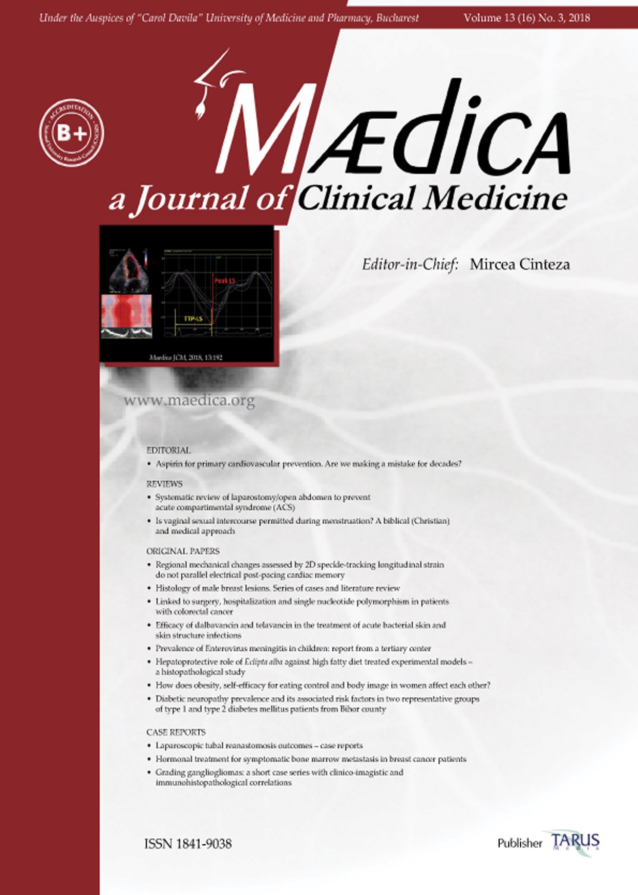
Regional Mechanical Changes Assessed by 2D Speckle-Tracking Longitudinal Strain do not Parallel Electrical Post-Pacing Cardiac Memory

SELECT ISSUE


MÆDICA - a Journal of Clinical Medicine | Vol. 13, nr. 3, 2018
CNCSIS - CMR - B+ OBBCSSR

Members Area


Regional Mechanical Changes Assessed by 2D Speckle-Tracking Longitudinal Strain do not Parallel Electrical Post-Pacing Cardiac Memory

Maria-Claudia-Berenice SURAN, Andrei-Dumitru MARGULESCU, Ramona BRUJA, Calin SILISTE and Dragos VINEREANU

ABSTRACT

Background. Cardiac memory (CM) refers to persistent T-wave changes that appear after cessation of a period of abnormal ventricular activation, such as ventricular pacing. Prior animal studies using tagged magnetic resonance imaging have suggested that CM is associated with prolonged action potential duration and increased strain of late-activated myocardial segments.
Objective. The aim of the present study is to determine whether CM induced by ventricular pacing in human subjects is accompanied by regional mechanical changes in late-activated myocardial segments, assessed by left ventricular (LV) longitudinal strain (peak LS) and time-to-peak longitudinal strain (TTP-LS), using 2D-speckle tracking echocardiography (2DSE).
Material and methods. We included 20 patients (16 women, age 71±11 years), with DDD pacemakers and with normal AV conduction and QRS/T morphology at baseline. CM was induced by DDD pacing with a short AV delay. ECGs and 2DSE were performed before pacing (baseline), at peak CM (immediately after two weeks of pacing), and at CM washout (4 weeks after cessation of pacing). We measured by echocardiography: left ventricular (LV) ejection fraction, LV diastolic function (E, A, E/E’), peak LS and TTP-LS for the earliest (i.e. adjacent to the pacing site) and latest (i.e. latest-activated during ventricular pacing) segments, using an 18-segment LV model.
Results. All patients had electrical (ECG) CM changes, which disappeared by CM washout. LV global systolic and diastolic functions, as well as regional LS (peak LS for both the latest and earliest activated segments) were similar between evaluations. TTP for the latest and earliest activated segments, as well as mean TTP-LS, increased from baseline to peak CM, but did not decrease at CM washout. The dispersion of TTP-LS was not changed.
Conclusion. These results suggest that regional mechanical changes, as can be assessed by 2DSE longitudinal strain, do not overlap electrical CM.
Keywords: 2-dimensional strain, 2DSE, echocardiography, cardiac memory.

Full text | PDF

Catecholaminergic Polymorphic Ventricular Tachycardia – Looking to the Future

SELECT ISSUE


MÆDICA - a Journal of Clinical Medicine | Vol. 12, nr. 4, 2017
CNCSIS - CMR - B+ OBBCSSR

Members Area


Catecholaminergic Polymorphic Ventricular Tachycardia – Looking to the Future

Andreea Elena VELCEA, Calin SILISTE and Dragos VINEREANU

ABSTRACT

Catecholaminergic polymorphic ventricular tachycardia (CPVT) is a rare inheritable cardiac disorder, characterized by polymorphic ventricular tachycardia (PVT) or bidirectional ventricular tachycardia, triggered by adrenergic stress, and manifested most frequently as syncope or sudden cardiac death. The disease has a heterogeneous genetic basis, with mutations in the genes encoding the ryanodine and calsequestrin channels accounting for the majority of cases.
The diagnosis of CPVT is established in individuals with polymorphic ventricular premature beats, PVT or bidirectional ventricular tachycardia documented during exercise or adrenergic stress, who have a structurally normal heart and normal resting ECG. Genetic testing completes the diagnosis, but is limited by the fact that, to date, about one third of cases are genotype-unknown.
Treatment strategies have improved as the knowledge of the disease has evolved, and several therapeutic options are now available. They include pharmacologic measures (especially non-selective beta-blockers and flecainide), but also more complex interventions, such implantation of internal cardiac defibrillators and left cardiac sympathetic denervation.
There are many unknowns to CPVT, but one that is essential to clinical practice is risk stratification, which will aid in a more targeted treatment of these patients. This goal is to be achieved by creating large patient registries and bio-banks, and ultimately by incorporating both clinical and genetic data into a risk stratification score.
Keywords: cathecolaminergic polymorphic ventricular tachycardia, bidirectional tachycardia, sudden cardiac death, genetic mutations

Full text | PDF

Left Ventricular Strain Analysis Reveals Better Synchrony and Diastolic Function for Septal Versus Apical Right Ventricular Permanent Pacing

SELECT ISSUE

MÆDICA - a Journal of Clinical Medicine | Vol. 9, nr. 3, 2014

CNCSIS - CMR - B+ OBBCSSR

Left Ventricular Strain Analysis Reveals Better Synchrony and Diastolic Function for Septal Versus Apical Right Ventricular Permanent Pacing

Roxana Cristina RIMBAS, Andrei Dumitru MARGULESCU, Calin SILISTE and Dragos VINEREANU

ABSTRACT

Objectives: Left ventricular function and synchrony may be altered by right ventricular (RV) apical pacing. Septal pacing might be a better alternative. We compared effects on cardiac synchrony and function, between the 2 pacing sites, in chronically implanted patients.

Material and methods: 40 pacing-dependent patients (74±9 years, 21 men), 20 paced at the apex, were studied 11±4 months after implantation (baseline); 32 of them were re-examined after 1 year. Systolic function was assessed from ejection fraction (EF), cardiac index (CI), mean longitudinal systolic strain (MLSS), and strain rate (MLSR); diastolic function from E/A, E/E’, and E/Vp ratios. Intraventricular dyssynchrony from standard deviation (SSD) and maximal difference (MAXS) of the 12 LV myocardial systolic timings, and sum of all times from the aortic valve closure to peak strain (SUMTAVC) for those segments with post-systolic shortening; interventricular synchrony from the aorto-pulmonary delay (APD).

Outcomes: Four patients died, all of them from the apical group. NYHA functional class was not different. Cardiac synchrony was not significantly different between the two pacing sites at baseline, and after 1 year follow-up. Although at baseline there was a greater dyssynchrony for the septal site, this did not progress at follow-up, whereas this increased for the apical site. Meanwhile, there was a higher LV filling pressure (E/E’ ratio) for the apical site at 1 year (13±6 vs.18±6; p=0.04).

Conclusions: Both septal and apical pacing sites affect negatively LV mechanical activation timings and synchrony. Apical, but not septal site, affects LV synchrony at 1 year, associated with increased filling pressure

Full text | PDF

Interdisciplinary Approach in a Complex Case of STEMI

SELECT ISSUE

MÆDICA - a Journal of Clinical Medicine | Vol. 9, nr. 4, 2014

CNCSIS - CMR - B+ OBBCSSR

Interdisciplinary Approach in a Complex Case of STEMI

Cristian A. UDROIU, Alexandru COTOBAN, Adrian URSULESCU, Calin SILISTE and Dragos VINEREANU

ABSTRACT

We reported the case of a young man with ST-Segment Elevation Myocardial Infarction (STEMI), with ventricular fibrillation on debut and cardiogenic shock, who needed a complex interdisciplinary approach for a favourable long term outcome.

A 43-year-old man was admitted with inferior STEMI and cardiogenic shock. First coronary angiography revealed total chronic occlusion of left anterior descending artery (LAD) and tight stenosis with thrombus on right coronary artery (RCA). Thrombus aspiration and stent implantation on RCA was performed with good results. LAD couldn’t be opened. Intraaortic balloon pump was implanted. Fortyeight hours later, we try again to open LAD, without success. After a lot of complications, all solved with difficulty, patient was discharged cachectic and with progressive exertion on mild exercise. Two months later an implantable cardioverter-defibrillator (ICD) was decided for persistent ventricular tachycardia and after one year he was referred to a cardiac surgery centre abroad for aneurismectomy with left ventricle (LV) reconstruction and mitral valve repair. The patient is currently asymptomatic with a normal social and professional life.

In conclusion, high performance cardiac surgery, after a complete interventional treatment, can improve quality of life and long-term outcome to a patient with severe cardiovascular disease.

Team work between clinical cardiologists, interventional cardiologists, electrophisyologists, intensivists and cardiac surgeons is the key to success.

Full text | PDF


plic-mail[email protected]

 Terms and conditions

© MÆDICA - a Journal of Clinical Medicine - All rights reserved